
Summary
Objectives To test the imaging performance and practical use of a novel dual-modality intravascular imaging system combining intravascular ultrasound (IVUS) and optical coherence tomography (OCT) into a single catheter.
Background IVUS enables assessing coronary plaque burden, a robust metric for patient prognosis, while OCT and OFDI provide high-resolution images of coronary microstructure and detailed assessment of stent implantation. Owing to their complementary strengths, co-registering IVUS and OFDI provides a more comprehensive assessment of coronary lesions during PCI.
Methods We developed a 2.6Fr imaging catheter integrating both IVUS and OFDI, interfacing to a dual-modality imaging console through a fast interchange connector. A novel algorithm fuses the IVUS and OFDI signals into a single combined image. We verified the performance of the two modalities and their visualization by imaging of cadaveric coronary arteries and tested practical imaging in the catheterization laboratory in swine in vivo.
Results Coronary atherosclerotic lesions of cadaver hearts revealed complementary optical and acoustic image features. Spatial co-registration of the modalities was confirmed by high correlation of measured lumen areas. Fused into a combined visualization, dual-modality imaging offers quantitative characterization of lesions, including plaque-burden and fibrous cap thickness. Imaging in vivo does not add procedural complexity compared to conventional single modality imaging.
Conclusion Dual-modality IVUS/OFDI imaging with fused visualization provides improved assessment of coronary atherosclerotic lesions and is compatible with a routine clinical setting. Combining the strength of the two modalities offers unique opportunities both as a powerful research instrument and to improve clinical management of patients undergoing PCI.

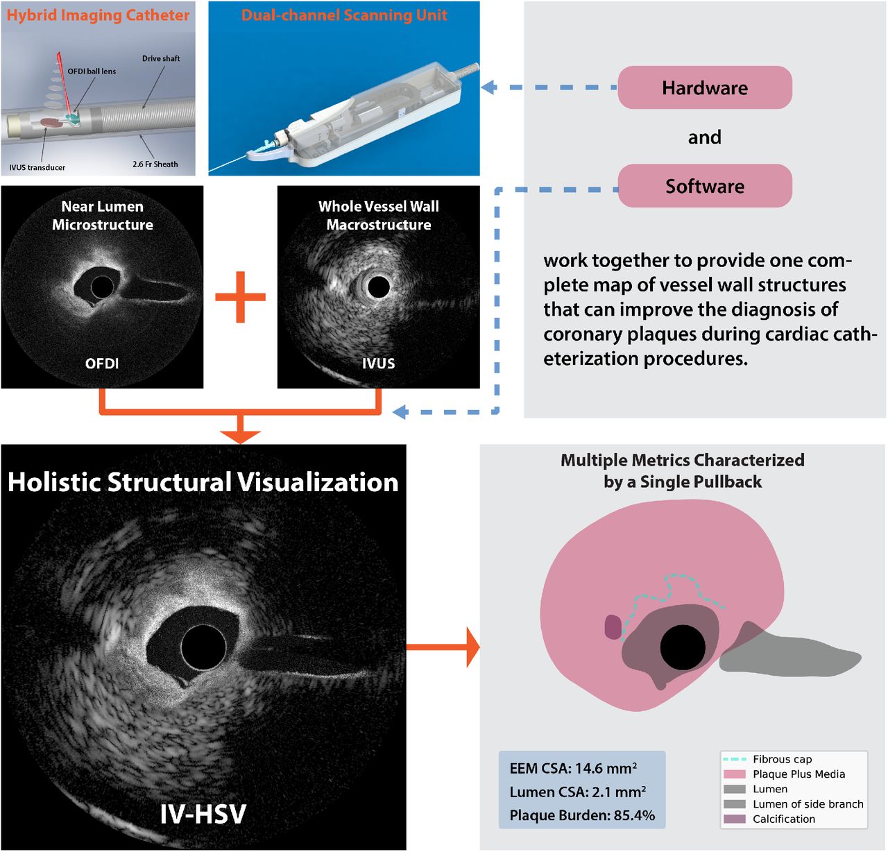
Visual abstract
Highlights
▪ A catheter-based imaging system integrating ultrasound and optical frequency domain imaging enables real time intravascular holistic structural visualization (IV-HSV) of coronary arterial lesions with a single pullback during cardiac catheterization procedures.
▪ A computational method to automatically fuse images from both modalities generates a single complete map of vessel wall structures - the IV-HSV image, which offers holistic investigation of coronary plaque key features, such as fibrous cap thickness, calcification, and plaque burden.
▪ The visualization of plaque morphology along the entire extent of the coronary arterial wall reveals vital information for guiding percutaneous coronary interventions and for advancing our understanding of the pathophysiology of coronary artery diseases.
Condensed abstract To facilitate a holistic investigation of coronary arterial lesions for both basic research and clinical interventions, this work presents a catheter-based imaging system integrating IVUS and OFDI, as well as a new rendering method that computationally fuses the intrinsically co-registered images from both modalities into a single cross-sectional map of vessel structures. Imaging human cadaveric coronary arteries shows the benefit of this system by revealing near-lumen microstructures and spatially correlated macrostructures deep inside the vessel wall. The co-registration accuracy and operation in a clinical setting of this system was demonstrated through swine catheterization in vivo.
Competing Interest Statement
Massachusetts General Hospital has patent licensing arrangements with Terumo Corporation. Drs. Bouma and Villiger have the right to receive royalties as part of the licensing arrangements. All other authors have reported that they have no relationships relevant to the contents of this paper to disclose.
Funding Statement
This work was supported by the National Institutes of Health (P41EB015903, K99AG059946, and R01HL119065) and by Terumo Corporation.
Author Declarations
I confirm all relevant ethical guidelines have been followed, and any necessary IRB and/or ethics committee approvals have been obtained.
Yes
The details of the IRB/oversight body that provided approval or exemption for the research described are given below:
The swine studies in this work were approved by the Massachusetts General Hospital Institutional Animal Care and Use Committee and performed with generally accepted guidelines governing such work.
All necessary patient/participant consent has been obtained and the appropriate institutional forms have been archived.
Yes
I understand that all clinical trials and any other prospective interventional studies must be registered with an ICMJE-approved registry, such as ClinicalTrials.gov. I confirm that any such study reported in the manuscript has been registered and the trial registration ID is provided (note: if posting a prospective study registered retrospectively, please provide a statement in the trial ID field explaining why the study was not registered in advance).
Yes
I have followed all appropriate research reporting guidelines and uploaded the relevant EQUATOR Network research reporting checklist(s) and other pertinent material as supplementary files, if applicable.
Yes
Footnotes
Financial support: This work was supported by the National Institutes of Health (P41EB015903, K99AG059946, and R01 HL119065) and by Terumo Corporation.
Relationship with industry: Massachusetts General Hospital has patent licensing arrangements with Terumo Corporation. Drs. Bouma and Villiger have the right to receive royalties as part of the licensing arrangements. All other authors have reported that they have no relationships relevant to the contents of this paper to disclose.
Abbreviations and Acronyms
- ACS
- acute coronary syndrome
- CSA
- cross sectional area
- EEM
- external elastic membrane
- IV-HSV
- intravascular holistic structural visualization
- IVUS
- intravascular ultrasound
- LAD
- left anterior descending artery
- LCX
- left circumflex artery
- OCT
- optical coherence tomography
- OFDI
- optical frequency domain imaging
- PCI
- percutaneous coronary intervention
- TCFA
- thin-capped fibroatheroma
The Chan Zuckerberg Initiative, Cold Spring Harbor Laboratory, the Sergey Brin Family Foundation, California Institute of Technology, Centre National de la Recherche Scientifique, Fred Hutchinson Cancer Center, Imperial College London, Massachusetts Institute of Technology, Stanford University, The University of Edinburgh, University of Washington, and Vrije Universiteit Amsterdam.


Subject Area
Reviews and Context
0
Comment
0
TRIP Peer Reviews
0
Community Reviews
0
Automated Services
0
Blogs/Media
Author Videos