
Diffusion Tensor Imaging Changes Do Not Affect Long-Term Neurodevelopment following Early Erythropoietin among Extremely Preterm Infants in the Preterm Erythropoietin Neuroprotection Trial

 , , , and
Abstract
:1. Introduction
2. Materials and Methods
2.1. Eligibility and Enrollment
2.2. Ethics
2.3. Imaging Protocol
2.4. DTI Data Processing
2.5. Neurodevelopmental Assessments
2.6. Statistical Analysis
2.7. Role of the Funding Source
3. Results
3.1. Enrollment and Group Demographics
3.2. Comparison of Adverse Events across Treatment Groups
3.3. Comparison of DTI Measures
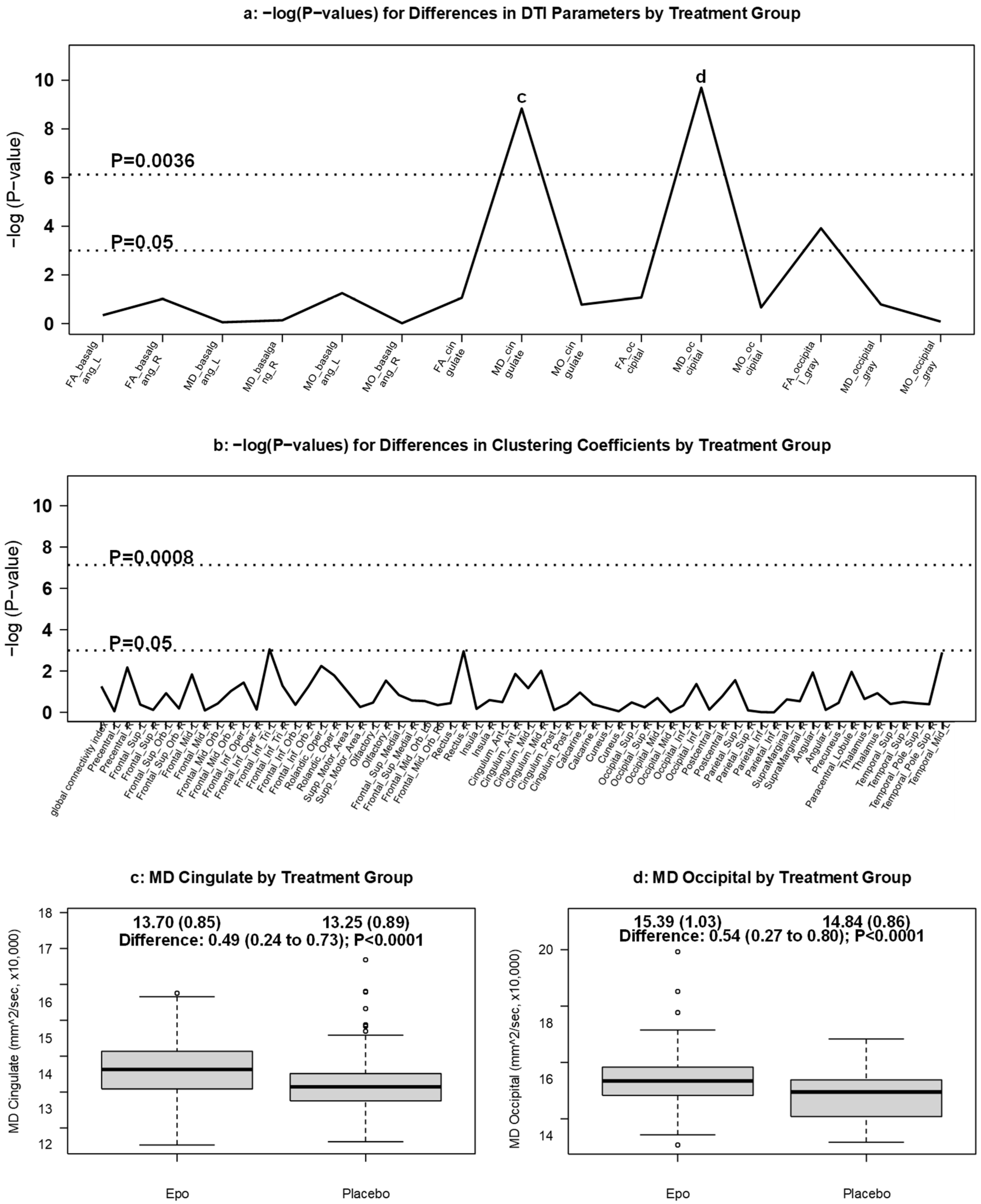
3.3.1. DTI Measures by Treatment Group
3.3.2. DTI Measures by Gestational Age
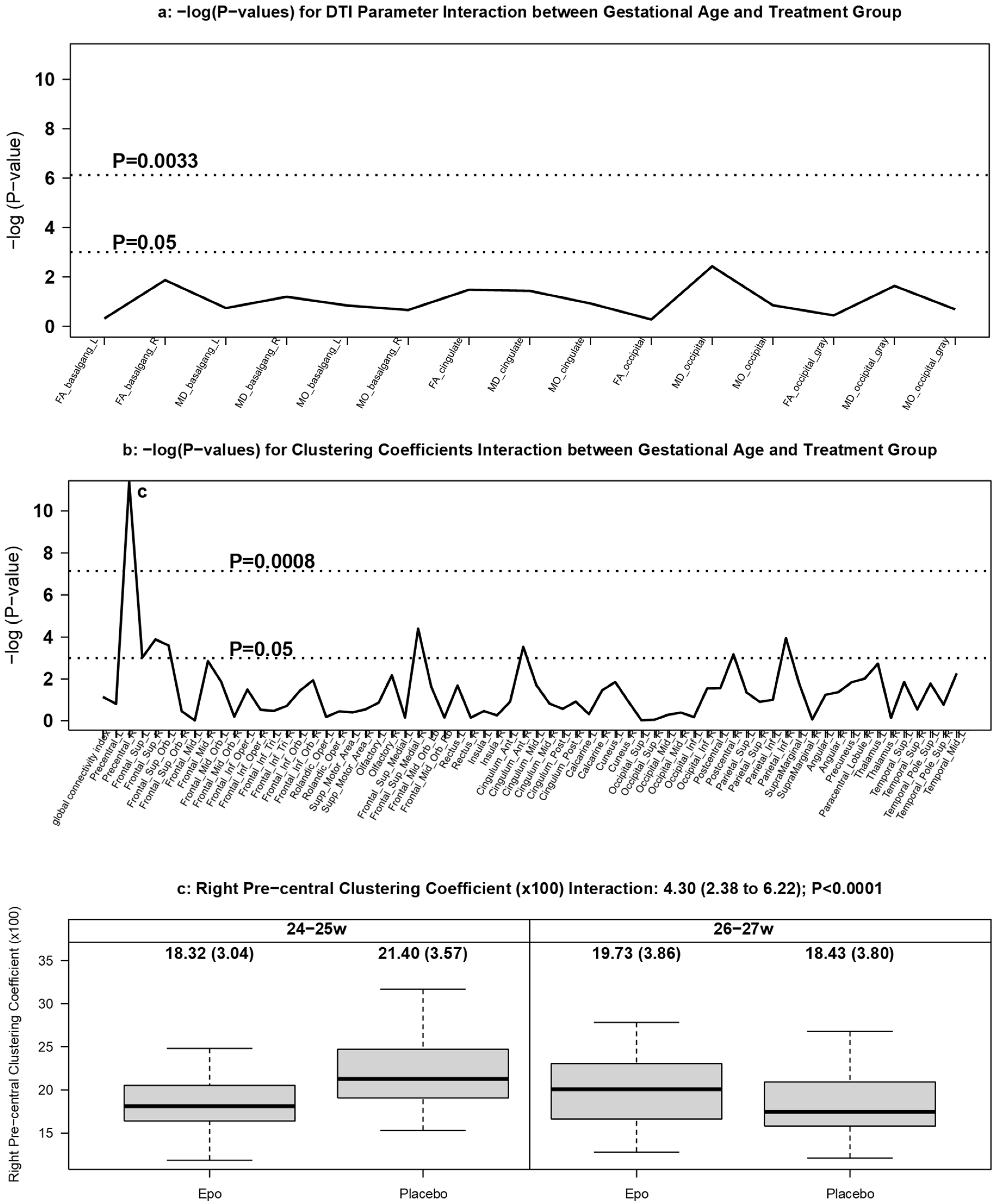
3.3.3. DTI Measures by GA and Treatment Group
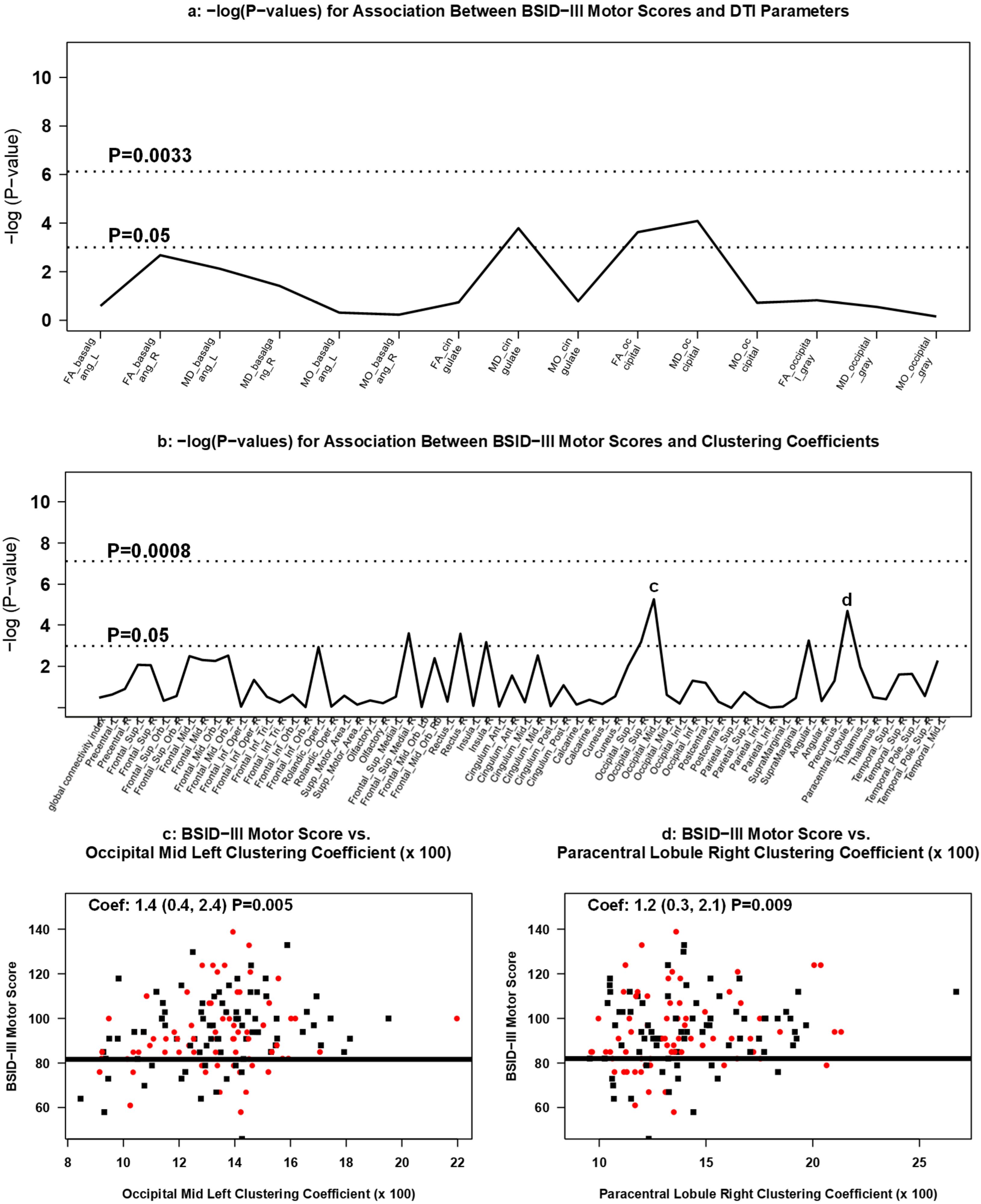
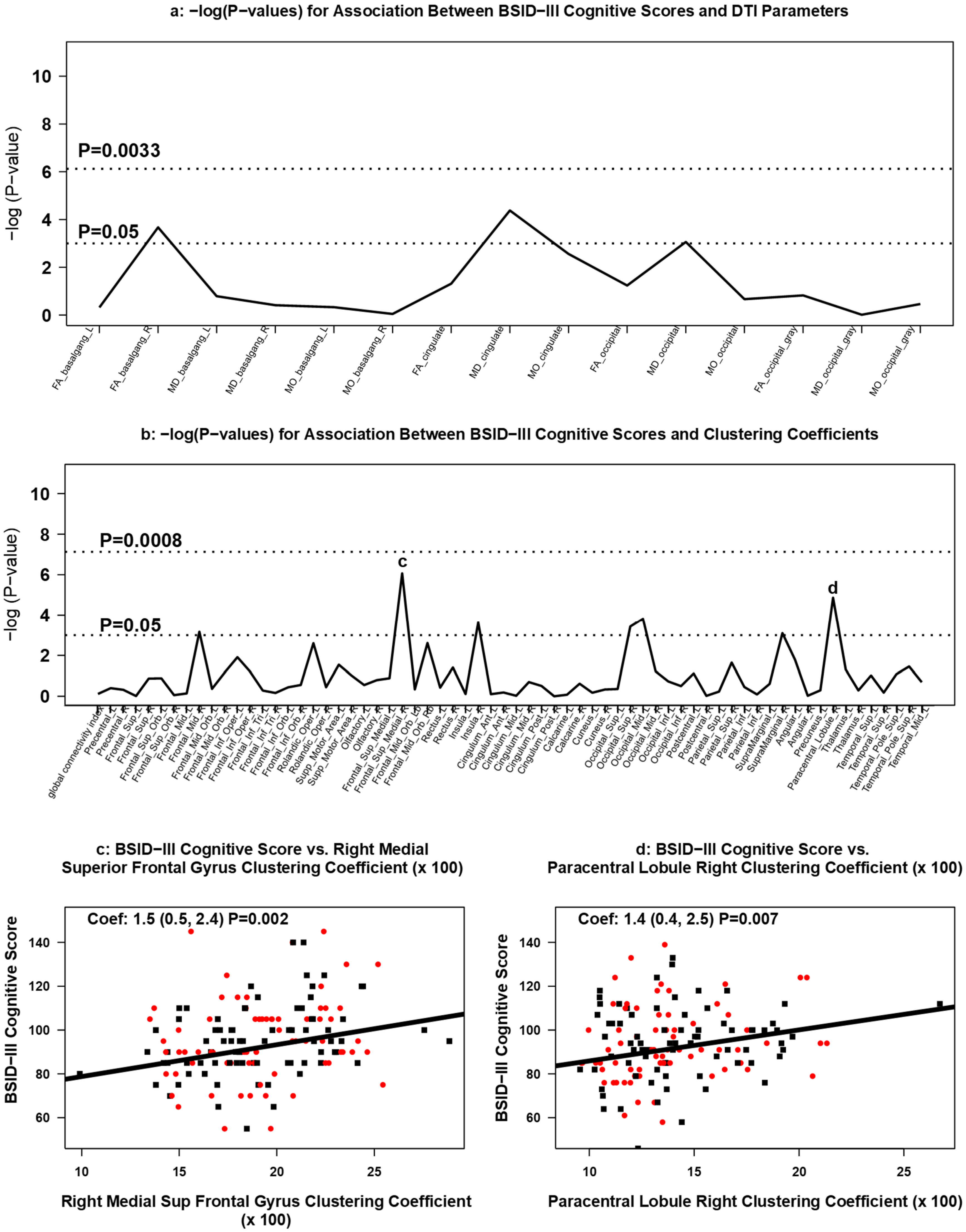
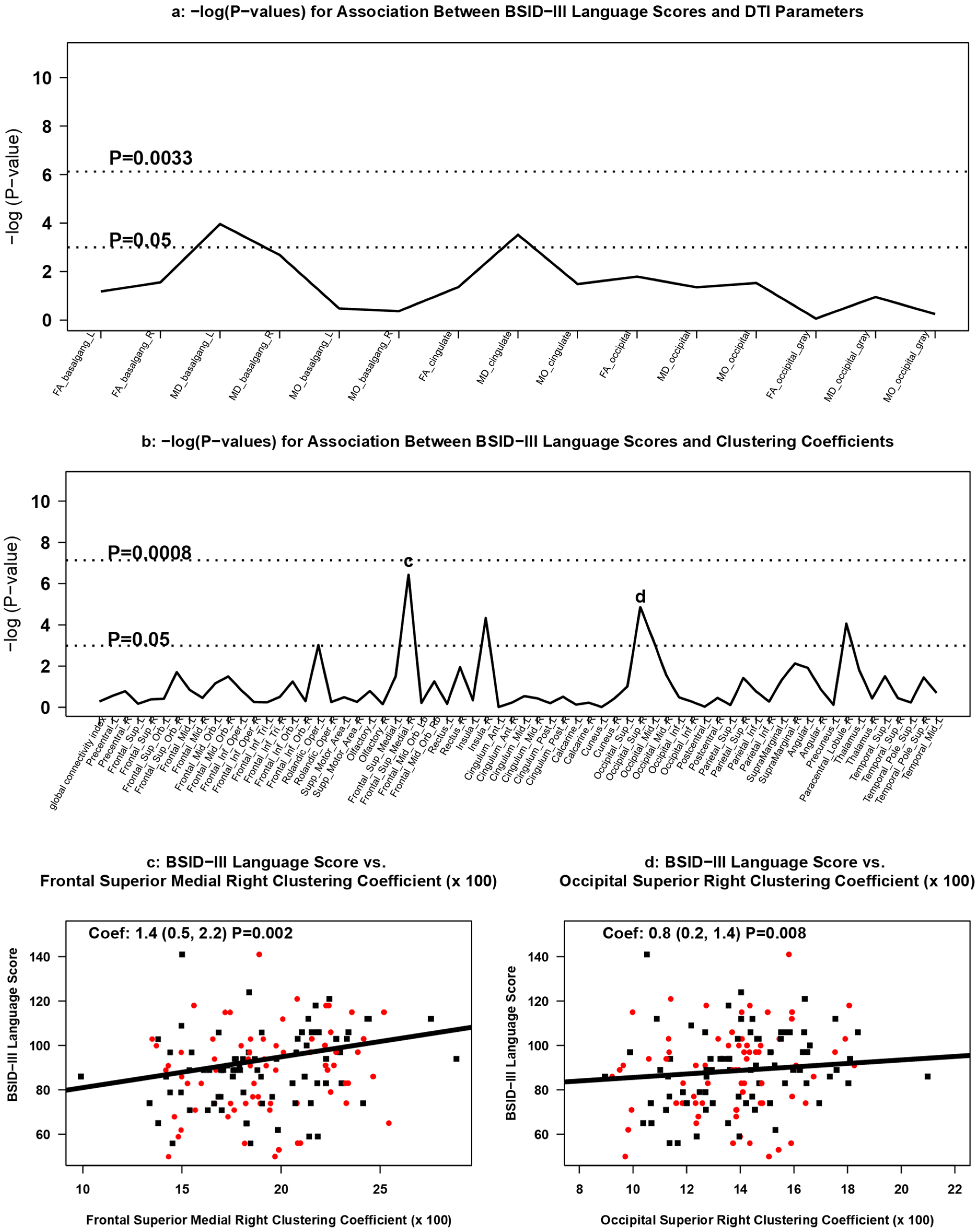
3.3.4. Association between DTI Measures and 2 Year Neurodevelopment
4. Discussion
5. Conclusions
Supplementary Materials
Author Contributions
Funding
Institutional Review Board Statement
Informed Consent Statement
Data Availability Statement
Acknowledgments
Conflicts of Interest
Appendix A
References
- VON. Vermont Oxford Network ELBW Follow-Up Report Birth Year 2008 All Centers; VON: Ottawa, ON, Canada, 2011. [Google Scholar]
- Younge, N.; Goldstein, R.F.; Bann, C.; Hintz, S.R.; Patel, R.; Smith, P.B.; Bell, E.; Rysavy, M.; Duncan, A.F.; Vohr, B.R.; et al. Survival and Neurodevelopmental Outcomes among Periviable Infants. N. Engl. J. Med. 2017, 376, 617–628. [Google Scholar] [CrossRef] [PubMed] [Green Version]
- Volpe, J.J. Brain injury in premature infants: A complex amalgam of destructive and developmental disturbances. Lancet Neurol. 2009, 8, 110–124. [Google Scholar] [CrossRef] [Green Version]
- Juul, S.E.; Comstock, B.A.; Wadhawan, R.; Mayock, D.E.; Courtney, S.E.; Robinson, T.; Ahmad, K.A.; Bendel-Stenzel, E.; Baserga, M.; LaGamma, E.F.; et al. A Randomized Trial of Erythropoietin for Neuroprotection in Preterm Infants. N. Engl. J. Med. 2020, 382, 233–243. [Google Scholar] [CrossRef] [PubMed]
- Stoll, B.J.; Hansen, N.I.; Bell, E.; Walsh, M.C.; Carlo, W.A.; Shankaran, S.; Laptook, A.R.; Sánchez, P.J.; Van Meurs, K.P.; Wyckoff, M.H.; et al. Trends in Care Practices, Morbidity, and Mortality of Extremely Preterm Neonates, 1993–2012. JAMA 2015, 314, 1039–1051. [Google Scholar] [CrossRef] [PubMed] [Green Version]
- Dyet, L.E.; Kennea, N.; Counsell, S.J.; Maalouf, E.F.; Ajayi-Obe, M.; Duggan, P.J.; Harrison, M.; Allsop, J.M.; Hajnal, J.; Herlihy, A.H.; et al. Natural History of Brain Lesions in Extremely Preterm Infants Studied with Serial Magnetic Resonance Imaging from Birth and Neurodevelopmental Assessment. Pediatrics 2006, 118, 536–548. [Google Scholar] [CrossRef]
- Inder, T.E.; Wells, S.J.; Mogridge, N.B.; Spencer, C.; Volpe, J.J. Defining the nature of the cerebral abnormalities in the premature infant: A qualitative magnetic resonance imaging study. J. Pediatrics 2003, 143, 171–179. [Google Scholar] [CrossRef]
- Patra, A.; Huang, H.; Bauer, J.A.; Giannone, P.J. Neurological consequences of systemic inflammation in the premature neonate. Neural Regen. Res. 2017, 12, 890–896. [Google Scholar] [CrossRef]
- Kocek, M.; Wilcox, R.; Crank, C.; Patra, K. Evaluation of the relationship between opioid exposure in extremely low birth weight infants in the neonatal intensive care unit and neurodevelopmental outcome at 2 years. Early Hum. Dev. 2016, 92, 29–32. [Google Scholar] [CrossRef]
- Woodward, L.J.; Clark, C.A.C.; Bora, S.; Inder, T.E. Neonatal White Matter Abnormalities an Important Predictor of Neurocognitive Outcome for Very Preterm Children. PLoS ONE 2012, 7, e51879. [Google Scholar] [CrossRef]
- Iwata, S.; Nakamura, T.; Hizume, E.; Kihara, H.; Takashima, S.; Matsuishi, T.; Iwata, O. Qualitative Brain MRI at Term and Cognitive Outcomes at 9 Years after Very Preterm Birth. Pediatrics 2012, 129, e1138–e1147. [Google Scholar] [CrossRef] [Green Version]
- Fukasawa, T.; Yamamoto, H.; Kubota, T. Diffusion tensor imaging at term-equivalent age in extremely-low-birth-weight infants with periventricular leukomalacia. No Hattatsu Brain Dev. 2012, 44, 19–24. [Google Scholar]
- Feldman, H.M.; Yeatman, J.D.; Lee, E.S.; Barde, L.H.F.; Gaman-Bean, S. Diffusion Tensor Imaging: A Review for Pediatric Researchers and Clinicians. J. Dev. Behav. Pediatrics 2010, 31, 346–356. [Google Scholar] [CrossRef] [Green Version]
- Ball, G.; Boardman, J.; Rueckert, D.; Aljabar, P.; Arichi, T.; Merchant, N.; Gousias, I.S.; Edwards, A.D.; Counsell, S.J. The Effect of Preterm Birth on Thalamic and Cortical Development. Cereb. Cortex 2011, 22, 1016–1024. [Google Scholar] [CrossRef]
- Ball, G.; Counsell, S.; Anjari, M.; Merchant, N.; Arichi, T.; Doria, V.; Rutherford, M.; Edwards, A.D.; Rueckert, D.; Boardman, J. An optimised tract-based spatial statistics protocol for neonates: Applications to prematurity and chronic lung disease. NeuroImage 2010, 53, 94–102. [Google Scholar] [CrossRef]
- Ball, G.; Srinivasan, L.; Aljabar, P.; Counsell, S.; Durighel, G.; Hajnal, J.; Rutherford, M.; Edwards, A.D. Development of cortical microstructure in the preterm human brain. Proc. Natl. Acad. Sci. USA 2013, 110, 9541–9546. [Google Scholar] [CrossRef] [Green Version]
- Eguíluz, V.M.; Chialvo, D.; Cecchi, G.A.; Baliki, M.; Apkarian, A.V. Scale-Free Brain Functional Networks. Phys. Rev. Lett. 2005, 94, 018102. [Google Scholar] [CrossRef] [PubMed] [Green Version]
- Xu, T.; Cullen, K.R.; Mueller, B.; Schreiner, M.W.; Lim, K.O.; Schulz, S.C.; Parhi, K.K. Network analysis of functional brain connectivity in borderline personality disorder using resting-state fMRI. NeuroImage Clin. 2016, 11, 302–315. [Google Scholar] [CrossRef] [PubMed] [Green Version]
- Honey, C.J.; Thivierge, J.-P.; Sporns, O. Can structure predict function in the human brain? NeuroImage 2010, 52, 766–776. [Google Scholar] [CrossRef] [PubMed]
- Jakab, A.; Ruegger, C.; Bucher, H.; Makki, M.; Hüppi, P.; Tuura, R.; Hagmann, C. Network based statistics reveals trophic and neuroprotective effect of early high dose erythropoetin on brain connectivity in very preterm infants. NeuroImage Clin. 2019, 22, 101806. [Google Scholar] [CrossRef] [PubMed]
- Ment, L.R.; Hirtz, D.; Hüppi, P. Imaging biomarkers of outcome in the developing preterm brain. Lancet Neurol. 2009, 8, 1042–1055. [Google Scholar] [CrossRef]
- Fischi-Gomez, E.; Vasung, L.; Meskaldji, D.-E.; Lazeyras, F.; Borradori-Tolsa, C.; Hagmann, P.; Barisnikov, K.; Thiran, J.-P.; Hüppi, P. Structural Brain Connectivity in School-Age Preterm Infants Provides Evidence for Impaired Networks Relevant for Higher Order Cognitive Skills and Social Cognition. Cereb. Cortex 2015, 25, 2793–2805. [Google Scholar] [CrossRef] [Green Version]
- Barnes-Davis, M.E.; Williamson, B.J.; Merhar, S.L.; Holland, S.K.; Kadis, D.S. Rewiring the extremely preterm brain: Altered structural connectivity relates to language function. NeuroImage Clin. 2020, 25, 102194. [Google Scholar] [CrossRef]
- Juul, S.E.; Mayock, D.E.; Comstock, B.A.; Heagerty, P.J. Neuroprotective potential of erythropoietin in neonates; design of a randomized trial. Matern. Health Neonatol. Perinatol. 2015, 1, 27. [Google Scholar] [CrossRef] [Green Version]
- Andersson, J.L.; Sotiropoulos, S. An integrated approach to correction for off-resonance effects and subject movement in diffusion MR imaging. NeuroImage 2016, 125, 1063–1078. [Google Scholar] [CrossRef] [Green Version]
- Andersson, J.L.R.; Graham, M.; Zsoldos, E.; Sotiropoulos, S. Incorporating outlier detection and replacement into a non-parametric framework for movement and distortion correction of diffusion MR images. NeuroImage 2016, 141, 556–572. [Google Scholar] [CrossRef] [PubMed] [Green Version]
- Jones, D.K.; Knösche, T.R.; Turner, R. White matter integrity, fiber count, and other fallacies: The do’s and don’ts of diffusion MRI. Neuroimage 2013, 73, 239–254. [Google Scholar] [CrossRef]
- Yoncheva, Y.N.; Somandepalli, K.; Reiss, P.T.; Kelly, C.; Di Martino, A.; Lazar, M.; Zhou, J.; Milham, M.P.; Castellanos, F.X. Mode of Anisotropy Reveals Global Diffusion Alterations in Attention-Deficit/Hyperactivity Disorder. J. Am. Acad. Child Adolesc. Psychiatry 2016, 55, 137–145. [Google Scholar] [CrossRef] [PubMed] [Green Version]
- Song, S.-K.; Sun, S.-W.; Ju, W.-K.; Lin, S.-J.; Cross, A.; Neufeld, A.H. Diffusion tensor imaging detects and differentiates axon and myelin degeneration in mouse optic nerve after retinal ischemia. NeuroImage 2003, 20, 1714–1722. [Google Scholar] [CrossRef] [PubMed]
- Song, S.-K.; Yoshino, J.; Le, T.Q.; Lin, S.-J.; Sun, S.-W.; Cross, A.; Armstrong, R.C. Demyelination increases radial diffusivity in corpus callosum of mouse brain. NeuroImage 2005, 26, 132–140. [Google Scholar] [CrossRef]
- Mori, R.; Khanna, R.; Pledge, D.; Nakayama, T. Meta-analysis of physiological effects of skin-to-skin contact for newborns and mothers. Pediatrics Int. 2010, 52, 161–170. [Google Scholar] [CrossRef]
- Smith, S.M.; Jenkinson, M.; Woolrich, M.W.; Beckmann, C.F.; Behrens, T.E.; Johansen-Berg, H.; Bannister, P.R.; De Luca, M.; Drobnjak, I.; Flitney, D.E.; et al. Advances in functional and structural MR image analysis and implementation as FSL. NeuroImage 2004, 23 (Suppl. 1), S208–S219. [Google Scholar] [CrossRef] [Green Version]
- Smith, S.M.; Jenkinson, M.; Johansen-Berg, H.; Rueckert, D.; Nichols, T.E.; Mackay, C.; Watkins, K.; Ciccarelli, O.; Cader, M.Z.; Matthews, P.M.; et al. Tract-based spatial statistics: Voxelwise analysis of multi-subject diffusion data. NeuroImage 2006, 31, 1487–1505. [Google Scholar] [CrossRef] [PubMed]
- Klein, A.; Andersson, J.; Ardekani, B.A.; Ashburner, J.; Avants, B.; Chiang, M.-C.; Christensen, G.E.; Collins, D.L.; Gee, J.; Hellier, P.; et al. Evaluation of 14 nonlinear deformation algorithms applied to human brain MRI registration. NeuroImage 2009, 46, 786–802. [Google Scholar] [CrossRef] [PubMed] [Green Version]
- Ewert, S.; Horn, A.; Finkel, F.; Li, N.; Kühn, A.A.; Herrington, T.M. Optimization and comparative evaluation of nonlinear deformation algorithms for atlas-based segmentation of DBS target nuclei. NeuroImage 2019, 184, 586–598. [Google Scholar] [CrossRef] [PubMed]
- Schwarz, C.G.; Reid, R.I.; Gunter, J.L.; Senjem, M.L.; Przybelski, S.A.; Zuk, S.M.; Whitwell, J.L.; Vemuri, P.; Josephs, K.A.; Kantarci, K.; et al. Improved DTI registration allows voxel-based analysis that outperforms Tract-Based Spatial Statistics. NeuroImage 2014, 94, 65–78. [Google Scholar] [CrossRef] [Green Version]
- Winkler, A.M.; Ridgway, G.R.; Webster, M.A.; Smith, S.; Nichols, T.E. Permutation inference for the general linear model. NeuroImage 2014, 92, 381–397. [Google Scholar] [CrossRef] [Green Version]
- Nichols, T.E.; Holmes, A.P. Nonparametric permutation tests for functional neuroimaging: A primer with examples. Hum. Brain Mapp. 2002, 15, 1–25. [Google Scholar] [CrossRef] [Green Version]
- Spisák, T.; Spisák, Z.; Zunhammer, M.; Bingel, U.; Smith, S.; Nichols, T.; Kincses, T. Probabilistic TFCE: A generalized combination of cluster size and voxel intensity to increase statistical power. NeuroImage 2019, 185, 12–26. [Google Scholar] [CrossRef]
- Richards, J.E.; Sanchez, C.; Phillips-Meek, M.; Xie, W. A database of age-appropriate average MRI templates. NeuroImage 2016, 124, 1254–1259. [Google Scholar] [CrossRef] [Green Version]
- Tzoutio-Mazoyera, N.; Landeau, B.; Papathanassiou, D.; Crivello, F.; Etard, O.; Delcroix, N.; Tzourio-Mazoyer, B.; Joliot, M. Automated Anatomical Labeling of Activations in SPM Using a Macroscopic Anatomical Parcellation of the MNI MRI Single-Subject Brain. NeuroImage 2002, 15, 273–289. [Google Scholar] [CrossRef]
- Watts, D.J.; Strogatz, S.H. Collective dynamics of ’small-world’ networks. Nature 1998, 393, 440–442. [Google Scholar] [CrossRef]
- Smyser, C.D.; Wheelock, M.D.; Limbrick, D.D.; Neil, J.J. Neonatal brain injury and aberrant connectivity. NeuroImage 2019, 185, 609–623. [Google Scholar] [CrossRef] [PubMed]
- Wee, C.-Y.; Tuan, T.A.; Broekman, B.F.P.; Ong, M.Y.; Chong, Y.S.; Kwek, K.; Shek, L.; Saw, S.-M.; Gluckman, P.D.; Fortier, M.V.; et al. Neonatal neural networks predict children behavioral profiles later in life. Hum. Brain Mapp. 2017, 38, 1362–1373. [Google Scholar] [CrossRef]
- Richards, T.L.; Berninger, V.W.; Yagle, K.; Abbott, R.D.; Peterson, D. Brain’s functional network clustering coefficient changes in response to instruction (RTI) in students with and without reading disabilities: Multi-leveled reading brain’s RTI. Cogent Psychol. 2018, 5, 5. [Google Scholar] [CrossRef] [PubMed]
- Tymofiyeva, O.; Hess, C.; Ziv, E.; Lee, P.N.; Glass, H.C.; Ferriero, D.M.; Barkovich, A.J.; Xu, D. A DTI-Based Template-Free Cortical Connectome Study of Brain Maturation. PLoS ONE 2013, 8, e63310. [Google Scholar] [CrossRef]
- Batalle, D.; Hughes, E.J.; Zhang, H.; Tournier, J.-D.; Tusor, N.; Aljabar, P.; Wali, L.; Alexander, D.C.; Hajnal, J.; Nosarti, C.; et al. Early development of structural networks and the impact of prematurity on brain connectivity. NeuroImage 2017, 149, 379–392. [Google Scholar] [CrossRef] [Green Version]
- Oishi, K.; Mori, S.; Donohue, P.K.; Ernst, T.; Anderson, L.; Buchthal, S.; Faria, A.; Jiang, H.; Li, X.; Miller, M.I.; et al. Multi-contrast human neonatal brain atlas: Application to normal neonate development analysis. NeuroImage 2011, 56, 8–20. [Google Scholar] [CrossRef] [PubMed] [Green Version]
- Rubinov, M.; Sporns, O. Complex network measures of brain connectivity: Uses and interpretations. NeuroImage 2010, 52, 1059–1069. [Google Scholar] [CrossRef] [PubMed]
- Oguz, I.; Farzinfar, M.; Matsui, J.; Budin, F.; Liu, Z.; Gerig, G.; Johnson, H.J.; Styner, M.A. DTIPrep: Quality control of diffusion-weighted images. Front. Aging Neurosci. 2014, 8, 4. [Google Scholar] [CrossRef] [Green Version]
- Wang, J.; Dong, Q.; Niu, H. The minimum resting-state fNIRS imaging duration for accurate and stable mapping of brain connectivity network in children. Sci. Rep. 2017, 7, 6461. [Google Scholar] [CrossRef] [PubMed] [Green Version]
- Xia, M.; Wang, J.; He, Y. BrainNet Viewer: A Network Visualization Tool for Human Brain Connectomics. PLoS ONE 2013, 8, e68910. [Google Scholar] [CrossRef] [Green Version]
- Diggle, P.; Diggle, P.J.; Heagerty, P.; Liang, K.Y.; Zeger, S. Analysis of Longitudinal Data, 2nd ed.; Oxford University Press: Oxford, UK, 2002. [Google Scholar]
- German, K.R.; Vu, P.T.; Comstock, B.A.; Ohls, R.K.; Heagerty, P.J.; Mayock, D.E.; Georgieff, M.; Rao, R.; Juul, S.E. Enteral Iron Supplementation in Infants Born Extremely Preterm and its Positive Correlation with Neurodevelopment; Post Hoc Analysis of the Preterm Erythropoietin Neuroprotection Trial Randomized Controlled Trial. J. Pediatrics 2021, in press. [Google Scholar] [CrossRef]
- Georgieff, M.K. The role of iron in neurodevelopment: Fetal iron deficiency and the developing hippocampus. Biochem. Soc. Trans. 2008, 36, 1267–1271. [Google Scholar] [CrossRef] [Green Version]
- Li, Y.; Juul, S.E.; Morris-Wiman, J.A.; A Calhoun, D.; Christensen, R.D. Erythropoietin Receptors Are Expressed in the Central Nervous System of Mid-Trimester Human Fetuses. Pediatric Res. 1996, 40, 376–380. [Google Scholar] [CrossRef] [Green Version]
- Liu, C.; Shen, K.; Liu, Z.; Noguchi, C.T. Regulated Human Erythropoietin Receptor Expression in Mouse Brain. J. Biol. Chem. 1997, 272, 32395–32400. [Google Scholar] [CrossRef] [PubMed] [Green Version]
- Juul, S.E.; Anderson, D.K.; Li, Y.; Christensen, R.D. Erythropoietin and Erythropoietin Receptor in the Developing Human Central Nervous System. Pediatric Res. 1998, 43, 40–49. [Google Scholar] [CrossRef] [Green Version]
- Dame, C.; Bartmann, P.; Wolber, E.-M.; Fahnenstich, H.; Hofmann, D.; Fandrey, J. Erythropoietin gene expression in different areas of the developing human central nervous system. Dev. Brain Res. 2000, 125, 69–74. [Google Scholar] [CrossRef]
- Shingo, T.; Sorokan, S.T.; Shimazaki, T.; Weiss, S. Erythropoietin regulates the in vitro and in vivo production of neuronal progenitors by mammalian forebrain neural stem cells. J. Neurosci. 2001, 21, 9733–9743. [Google Scholar] [CrossRef] [PubMed] [Green Version]
- Juul, S.E.; Pet, G.C. Erythropoietin and Neonatal Neuroprotection. Clin. Perinatol. 2015, 42, 469–481. [Google Scholar] [CrossRef] [PubMed] [Green Version]
- van der Kooij, M.A.; Groenendaal, F.; Kavelaars, A.; Heijnen, C.J.; van Bel, F. Neuroprotective properties and mechanisms of erythropoietin in in vitro and in vivo experimental models for hypoxia/ischemia. Brain Res. Rev. 2008, 59, 22–33. [Google Scholar] [CrossRef] [PubMed]
- Kumral, A.; Gonenc, S.; Açikgoz, O.; Sonmez, A.; Genc, K.K.; Yilmaz, O.; Gökmen, N.; Duman, N.; Ozkan, H. Erythropoietin Increases Glutathione Peroxidase Enzyme Activity and Decreases Lipid Peroxidation Levels in Hypoxic-Ischemic Brain Injury in Neonatal Rats. Neonatology 2005, 87, 15–18. [Google Scholar] [CrossRef] [PubMed]
- Wang, L.; Zhang, Z.; Wang, Y.; Zhang, R.; Chopp, M. Treatment of Stroke With Erythropoietin Enhances Neurogenesis and Angiogenesis and Improves Neurological Function in Rats. Stroke 2004, 35, 1732–1737. [Google Scholar] [CrossRef] [PubMed] [Green Version]
- Iwai, M.; Cao, G.; Yin, W.; Stetler, R.A.; Liu, J.; Chen, J. Erythropoietin Promotes Neuronal Replacement through Revascularization and Neurogenesis after Neonatal Hypoxia/Ischemia in Rats. Stroke 2007, 38, 2795–2803. [Google Scholar] [CrossRef] [PubMed] [Green Version]
- Osredkar, D.; Sall, J.W.; Bickler, P.E.; Ferriero, D.M. Erythropoietin promotes hippocampal neurogenesis in in vitro models of neonatal stroke. Neurobiol. Dis. 2010, 38, 259–265. [Google Scholar] [CrossRef] [Green Version]
- Wassink, G.; Davidson, J.O.; Dhillon, S.K.; Fraser, M.; Galinsky, R.; Bennet, L.; Gunn, A.J. Partial white and grey matter protection with prolonged infusion of recombinant human erythropoietin after asphyxia in preterm fetal sheep. J. Cereb. Blood Flow Metab. 2017, 37, 1080–1094. [Google Scholar] [CrossRef] [Green Version]
- Kerendi, F.; Halkos, M.E.; Kin, H.; Corvera, J.S.; Brat, D.J.; Wagner, M.B.; Vinten-Johansen, J.; Zhao, Z.-Q.; Forbess, J.M.; Kanter, K.R.; et al. Upregulation of hypoxia inducible factor is associated with attenuation of neuronal injury in neonatal piglets undergoing deep hypothermic circulatory arrest. J. Thorac. Cardiovasc. Surg. 2005, 130, 1079.e1–1079.e10. [Google Scholar] [CrossRef] [Green Version]
- Traudt, C.M.; McPherson, R.J.; Bauer, L.A.; Richards, T.L.; Burbacher, T.M.; McAdams, R.M.; Juul, S.E. Concurrent Erythropoietin and Hypothermia Treatment Improve Outcomes in a Term Nonhuman Primate Model of Perinatal Asphyxia. Dev. Neurosci. 2013, 35, 491–503. [Google Scholar] [CrossRef] [Green Version]
- Geng, F.; Mai, X.; Zhan, J.; Xu, L.; Zhao, Z.; Georgieff, M.; Shao, J.; Lozoff, B. Impact of Fetal-Neonatal Iron Deficiency on Recognition Memory at 2 Months of Age. J. Pediatrics 2015, 167, 1226–1232. [Google Scholar] [CrossRef] [Green Version]
- Puia-Dumitrescu, M.; Comstock, B.A.; Li, S.; Heagerty, P.J.; Perez, K.M.; Law, J.B.; Wood, T.R.; Gogcu, S.; Mayock, D.E.; Juul, S.E.; et al. Assessment of 2-Year Neurodevelopmental Outcomes in Extremely Preterm Infants Receiving Opioids and Benzodiazepines. JAMA Netw. Open 2021, 4, e2115998. [Google Scholar] [CrossRef]
- O’Gorman, R.L.; Bucher, H.U.; Held, U.; Koller, B.M.; Hüppi, P.S.; Hagmann, C.F.; Swiss EPO Neuroprotection Trial Group. Tract-based spatial statistics to assess the neuroprotective effect of early erythropoietin on white matter development in preterm infants. Brain 2015, 138, 388–397. [Google Scholar] [CrossRef] [Green Version]
- Yang, S.S.; Xu, F.L.; Cheng, H.Q.; Xu, H.R.; Yang, L.; Xing, J.Y.; Cheng, L. Effect of early application of recombinant human erythropoietin on white matter development in preterm infants. Zhongguo Dang Dai Er Ke Za Zhi 2018, 20, 346–351. [Google Scholar] [PubMed]
- Phillips, J.; Yeo, R.A.; Caprihan, A.; Cannon, D.C.; Patel, S.; Winter, S.; Steffen, M.; Campbell, R.; Wiedmeier, S.; Baker, S.; et al. Neuroimaging in former preterm children who received erythropoiesis stimulating agents. Pediatric Res. 2017, 82, 685–690. [Google Scholar] [CrossRef] [Green Version]
- Rao, R.; Tkac, I.; Unger, E.L.; Ennis, K.; Hurst, A.; Schallert, T.; Connor, J.; Felt, B.; Georgieff, M.K. Iron supplementation dose for perinatal iron deficiency differentially alters the neurochemistry of the frontal cortex and hippocampus in adult rats. Pediatric Res. 2012, 73, 31–37. [Google Scholar] [CrossRef] [PubMed] [Green Version]
- Buonocore, G.; Perrone, S.; Bracci, R. Free Radicals and Brain Damage in the Newborn. Biol. Neonate 2001, 79, 180–186. [Google Scholar] [CrossRef]
- Wang, Y.; Wu, Y.; Li, T.; Wang, X.; Zhu, C. Iron Metabolism and Brain Development in Premature Infants. Front. Physiol. 2019, 10, 463. [Google Scholar] [CrossRef] [PubMed]
- Khwaja, O.; Volpe, J.J. Pathogenesis of cerebral white matter injury of prematurity. Arch. Dis. Child. Fetal Neonatal Ed. 2008, 93, F153–F161. [Google Scholar] [CrossRef]
- Juul, S.E.; Vu, P.T.; Comstock, B.A.; Wadhawan, R.; Mayock, D.E.; Courtney, S.E.; Robinson, T.; Ahmad, K.A.; Bendel-Stenzel, E.; Baserga, M.; et al. Effect of High-Dose Erythropoietin on Blood Transfusions in Extremely Low Gestational Age Neonates: Post Hoc Analysis of a Randomized Clinical Trial. JAMA Pediatrics 2020, 174, 933–943. [Google Scholar] [CrossRef]
- Pogribna, U.; Yu, X.; Burson, K.; Zhou, Y.; Lasky, R.E.; Narayana, P.A.; Parikh, N.A. Perinatal Clinical Antecedents of White Matter Microstructural Abnormalities on Diffusion Tensor Imaging in Extremely Preterm Infants. PLoS ONE 2013, 8, e72974. [Google Scholar] [CrossRef] [Green Version]
- Anjari, M.; Srinivasan, L.; Allsop, J.M.; Hajnal, J.; Rutherford, M.; Edwards, A.D.; Counsell, S. Diffusion tensor imaging with tract-based spatial statistics reveals local white matter abnormalities in preterm infants. NeuroImage 2007, 35, 1021–1027. [Google Scholar] [CrossRef]
- Thompson, D.K.; Kelly, C.; Chen, J.; Beare, R.; Alexander, B.; Seal, M.; Lee, K.J.; Matthews, L.G.; Anderson, P.J.; Doyle, L.W.; et al. Characterisation of brain volume and microstructure at term-equivalent age in infants born across the gestational age spectrum. NeuroImage Clin. 2019, 21, 101630. [Google Scholar] [CrossRef]
- Skiöld, B.; Horsch, S.; Hallberg, B.; Engström, M.; Nagy, Z.; Mosskin, M.; Blennow, M.; Ådén, U. White matter changes in extremely preterm infants, a population-based diffusion tensor imaging study. Acta Paediatr. 2010, 99, 842–849. [Google Scholar] [CrossRef]
- Rose, J.; Vassar, R.; Cahill-Rowley, K.; Guzman, X.S.; Hintz, S.R.; Stevenson, D.K.; Barnea-Goraly, N. Neonatal physiological correlates of near-term brain development on MRI and DTI in very-low-birth-weight preterm infants. NeuroImage Clin. 2014, 5, 169–177. [Google Scholar] [CrossRef] [PubMed] [Green Version]
- Kelly, C.E.; Thompson, D.K.; Cheong, J.L.; Chen, J.; Olsen, J.E.; Eeles, A.L.; Walsh, J.M.; Seal, M.L.; Anderson, P.J.; Doyle, L.W.; et al. Brain structure and neurological and behavioural functioning in infants born preterm. Dev. Med. Child Neurol. 2019, 61, 820–831. [Google Scholar] [CrossRef] [PubMed] [Green Version]
- Pogribna, U.; Burson, K.; Lasky, R.E.; Narayana, P.A.; Evans, P.W.; Parikh, N.A. Role of diffusion tensor imaging as an independent predictor of cognitive and language development in extremely low-birth-weight infants. Am. J. Neuroradiol. 2013, 35, 790–796. [Google Scholar] [CrossRef] [PubMed] [Green Version]
- Anderson, P.; Burnett, A. Assessing developmental delay in early childhood—Concerns with the Bayley-III scales. Clin. Neuropsychol. 2017, 31, 371–381. [Google Scholar] [CrossRef]
- Flynn, R.S.; Huber, M.D.; DeMauro, S.B. Predictive Value of the BSID-II and the Bayley-III for Early School Age Cognitive Function in Very Preterm Infants. Glob. Pediatric Health 2020, 7, 2333794X20973146. [Google Scholar] [CrossRef]
- Sharp, M.; DeMauro, S.B. Counterbalanced Comparison of the BSID-II and Bayley-III at Eighteen to Twenty-two Months Corrected Age. J. Dev. Behav. Pediatrics 2017, 38, 322–329. [Google Scholar] [CrossRef]
- Renier, L.A.; Anurova, I.; De Volder, A.G.; Carlson, S.; VanMeter, J.; Rauschecker, J.P. Preserved Functional Specialization for Spatial Processing in the Middle Occipital Gyrus of the Early Blind. Neuron 2010, 68, 138–148. [Google Scholar] [CrossRef] [Green Version]
- Spasojević, G.; Malobabic, S.; Pilipović-Spasojević, O.; Djukić-Macut, N.; Maliković, A. Morphology and digitally aided morphometry of the human paracentral lobule. Folia Morphol. 2013, 72, 10–16. [Google Scholar] [CrossRef] [Green Version]
- Japee, S.; Holiday, K.; Satyshur, M.D.; Mukai, I.; Ungerleider, L.G. A role of right middle frontal gyrus in reorienting of attention: A case study. Front. Syst. Neurosci. 2015, 9, 23. [Google Scholar] [CrossRef] [Green Version]
- Zhang, S.; Tsai, S.-J.; Hu, S.; Xu, J.; Chao, H.H.; Calhoun, V.D.; Li, C.-S.R. Independent component analysis of functional networks for response inhibition: Inter-subject variation in stop signal reaction time. Hum. Brain Mapp. 2015, 36, 3289–3302. [Google Scholar] [CrossRef] [PubMed] [Green Version]
- Sughrue, M.E. Butterfly Glioma Resection: Surgery around the Initiation Axis, in New Techniques for Management of ’Inoperable’ Gliomas; Michael, I.Y., Sughrue, E., Eds.; Academic Press: Cambridge, MA, USA, 2019; pp. 103–115. [Google Scholar]
- Lane, C.; Kanjlia, S.; Omaki, A.; Bedny, M. “Visual” Cortex of Congenitally Blind Adults Responds to Syntactic Movement. J. Neurosci. 2015, 35, 12859–12868. [Google Scholar] [CrossRef] [PubMed] [Green Version]
- Lin, F.-H.; Chen, Y.-J.; Belliveau, J.W.; Wald, L.L. A wavelet-based approximation of surface coil sensitivity profiles for correction of image intensity inhomogeneity and parallel imaging reconstruction. Hum. Brain Mapp. 2003, 19, 96–111. [Google Scholar] [CrossRef] [PubMed]
- Wilson, S.; Pietsch, M.; Cordero-Grande, L.; Price, A.N.; Hutter, J.; Xiao, J.; McCabe, L.; Rutherford, M.A.; Hughes, E.J.; Counsell, S.J.; et al. Development of human white matter pathways in utero over the second and third trimester. Proc. Natl. Acad. Sci. USA 2021, 118, e2023598118. [Google Scholar] [CrossRef] [PubMed]


| MRI Cohort | Non-MRI Cohort * | |||
|---|---|---|---|---|
| Placebo | Epo | Overall | ||
| Maternal demographics, N (%) | N = 101 | N = 93 | N = 194 | N = 228 |
| Age, mean (SD) | 27.9 (6.2) | 28.9 (6.6) | 28.4 (6.4) | 29.1 (6.2) |
| Race | ||||
| Hispanic | 38 (38%) | 27 (29%) | 65 (34%) | 51 (22%) ** |
| White | 62 (61%) | 64 (69%) | 126 (65%) | 134 (59%) |
| Black | 26 (26%) | 23 (25%) | 49 (25%) | 77 (34%) |
| Other/Not reported | 13 (13%) | 6 (6%) | 19 (10%) | 17 (7.5%) |
| Education | ||||
| High School or less | 45 (45%) | 26 (28%) | 71 (37%) | 75 (33%) |
| Some college | 23 (23%) | 26 (28%) | 49 (25%) | 81 (36%) |
| College degree or greater | 23 (23%) | 25 (27%) | 48 (25%) | 49 (21%) |
| Not reported | 10 (10%) | 16 (17%) | 26 (13%) | 23 (11%) |
| Neonatal data at enrollment, N (%) | ||||
| Delivery complications | 10 (10%) | 13 (14%) | 23 (12%) | 33 (14%) |
| Antenatal steroids | 92 (91%) | 83 (89%) | 175 (90%) | 207 (91%) |
| Chorioamnionitis | 16 (16%) | 16 (17%) | 32 (16%) | 33 (14%) |
| Caesarean delivery | 65 (64%) | 60 (65%) | 125 (64%) | 161 (71%) |
| Delayed cord clamping | 44 (58%) | 49 (61%) | 93 (60%) | 67 (38%) *** |
| Female | 56 (55%) | 41 (44%) | 97 (50%) | 103 (45%) |
| Gestational age | ||||
| 24 weeks | 25 (25%) | 15 (16%) | 40 (20%) | 57 (25%) |
| 25 weeks | 24 (24%) | 23 (25%) | 47 (24%) | 56 (25%) |
| 26 weeks | 26 (26%) | 27 (29%) | 53 (28%) | 58 (25%) |
| 27 weeks | 26 (26%) | 28 (30%) | 54 (28%) | 57 (25%) |
| Mean (SD) | 25.9 (1.2) | 26.1 (1.1) | 26.0 (1.1) | 25.9 (1.1) |
| Multiple gestation | 19 (19%) | 24 (26%) | 43 (22%) | 60 (26%) |
| Infant weight (grams), mean (SD) | 805.2 (176.3) | 859.7 (177.6) | 831.3 (178.6) | 783.1 (183.4) *** |
| Apgar score at 5 min, mean (SD) | 6.5 (1.8) | 6.6 (1.9) | 6.6 (1.9) | 6.0 (2.1) *** |
| Epo level at birth, median (IQR) | N = 85 7.1 (4.2, 14.5) | N = 74 8.5 (4.8, 49.3) | N = 159 7.3 (4.4, 22.7) | N = 185 8.4 (4.2, 24.8) |
| MRI Cohort | Non-MRI Cohort * | |||
|---|---|---|---|---|
| Placebo | Epo | Overall | ||
| Postnatal markers of instability, N (%) | N = 101 | N = 93 | N = 194 | N = 228 |
| Necrotizing Enterocolitis (NEC) | 6 (5.9%) | 2 (2.2%) | 8 (4.1%) | 15 (6.6%) |
| Spontaneous Intestinal Perforation (SIP) | 2 (2.0%) | 1 (1.1%) | 3 (1.5%) | 11 (4.8%) |
| Sepsis | 3 (3.0%) | 3 (3.2%) | 6 (3.1%) | 28 (12%) ** |
| Retinopathy of Prematurity (ROP) | 8 (7.9%) | 6 (6.5%) | 14 (7.2%) | 19 (8.3%) |
| Severe Intraventricular hemorrhage (IVH) | 4 (5.9%) | 2 (2.2%) | 6 (3.1%) | 36 (16%) § |
| Risk factors for NDI, N (%) | ||||
| Lowest ferritin in ng/mL (any time) | ||||
| <76 | 22/96 (23%) | 61/89 (69%) | 83/185 (45%) | 75/200 (38%) |
| <40 | 6/96 (6.3%) | 39/89 (44%) | 45/185 (24%) | 40/200 (20%) |
| Chronic lung disease (CLD) | 42 (42%) | 28 (30%) | 70 (36%) | 86 (38%) |
| Outcomes at Age 2, mean (SD) | N = 81 | N = 73 | N = 154 | N = 184 |
| BSID-III Cognitive | 95.1 (15.8) | 95.7 (18.6) | 95.4 (17.2) | 87.4 (16.1) § |
| BSID-III Motor | 94.2 (15.9) | 93.4 (16.7) | 93.8 (16.2) | 85.7 (17.4) § |
| BSID-III Language | 89.8 (16.7) | 88.2 (19.0) | 89.0 (17.8) | 85.7 (18.2) |
Publisher’s Note: MDPI stays neutral with regard to jurisdictional claims in published maps and institutional affiliations. |
© 2021 by the authors. Licensee MDPI, Basel, Switzerland. This article is an open access article distributed under the terms and conditions of the Creative Commons Attribution (CC BY) license (https://creativecommons.org/licenses/by/4.0/).
Share and Cite
Law, J.B.; Comstock, B.A.; Richards, T.L.; Traudt, C.M.; Wood, T.R.; Mayock, D.E.; Heagerty, P.J.; Juul, S.E., on behalf of the PENUT Trial Consortium. Diffusion Tensor Imaging Changes Do Not Affect Long-Term Neurodevelopment following Early Erythropoietin among Extremely Preterm Infants in the Preterm Erythropoietin Neuroprotection Trial. Brain Sci. 2021, 11, 1360. https://doi.org/10.3390/brainsci11101360
Law JB, Comstock BA, Richards TL, Traudt CM, Wood TR, Mayock DE, Heagerty PJ, Juul SE on behalf of the PENUT Trial Consortium. Diffusion Tensor Imaging Changes Do Not Affect Long-Term Neurodevelopment following Early Erythropoietin among Extremely Preterm Infants in the Preterm Erythropoietin Neuroprotection Trial. Brain Sciences. 2021; 11(10):1360. https://doi.org/10.3390/brainsci11101360
Chicago/Turabian StyleLaw, Janessa B., Bryan A. Comstock, Todd L. Richards, Christopher M. Traudt, Thomas R. Wood, Dennis E. Mayock, Patrick J. Heagerty, and Sandra E. Juul on behalf of the PENUT Trial Consortium. 2021. "Diffusion Tensor Imaging Changes Do Not Affect Long-Term Neurodevelopment following Early Erythropoietin among Extremely Preterm Infants in the Preterm Erythropoietin Neuroprotection Trial" Brain Sciences 11, no. 10: 1360. https://doi.org/10.3390/brainsci11101360
APA StyleLaw, J. B., Comstock, B. A., Richards, T. L., Traudt, C. M., Wood, T. R., Mayock, D. E., Heagerty, P. J., & Juul, S. E., on behalf of the PENUT Trial Consortium. (2021). Diffusion Tensor Imaging Changes Do Not Affect Long-Term Neurodevelopment following Early Erythropoietin among Extremely Preterm Infants in the Preterm Erythropoietin Neuroprotection Trial. Brain Sciences, 11(10), 1360. https://doi.org/10.3390/brainsci11101360